Quick Answer
Shockwave therapy is a non-invasive treatment podiatrists use for persistent heel pain, especially long-standing plantar fasciitis and some cases of Achilles tendinopathy. It works by delivering controlled acoustic waves into the sore tissue to stimulate healing and calm long-term irritation.
It is most often recommended when heel pain has been present for several months and has not improved with standard care. Standard care usually includes stretching, supportive footwear, orthotics, and changes to activity levels.
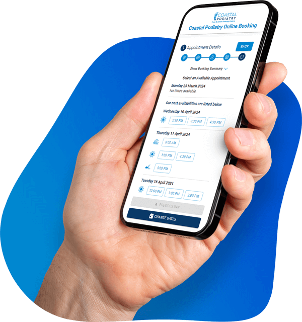
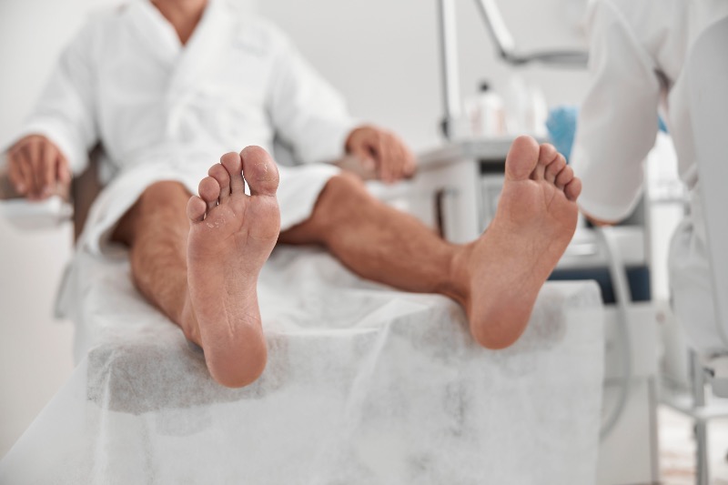
In an appointment, a podiatrist uses a handheld device to deliver short pulses to the tender area of the heel. A session usually takes about 10 to 20 minutes, and many patients need a small course of treatments over several weeks.
Most people can walk and continue daily life as normal during treatment, although your podiatrist may suggest reducing running or jumping temporarily. The aim is steady improvement, with pain typically easing gradually as the tissue repairs.
Understanding Heel Pain: Why It Is Such a Common Foot Problem
Heel pain is one of the most common reasons people book a podiatry appointment. The heel takes a large share of the load every time your foot hits the ground, so it is exposed to repeated impact and strain across the day.
The tissues around the heel are designed to absorb shock and support the arch, but they can be overwhelmed. If the load on the foot rises faster than the tissue can adapt, tiny injuries can develop and become painful.
Common reasons heel pain shows up in everyday life include:
- long hours standing on hard floors
- sudden increases in walking, running, or sport
- reduced calf flexibility
- worn or unsupportive shoes
- changes in weight, training, or work demands
Because you cannot fully “rest” your feet in the way you might rest a shoulder or elbow, heel pain can linger. That is why some people need a more targeted treatment plan, and why shockwave therapy is sometimes considered.
The Role of the Heel in Walking and Movement
The heel bone, called the calcaneus, is the largest bone in the foot. It works like a base plate for several important structures, and it takes a strong impact at heel strike in walking and running.
The plantar fascia, Achilles tendon, and several stabilising tissues connect to or pass around the heel. When those tissues repeatedly absorb force, they can become irritated, especially if mechanics or footwear increase the load.
Key structures commonly involved in heel pain include:
- plantar fascia (under the foot)
- Achilles tendon (back of the heel)
- heel fat pad (cushion under the heel)
- small nerves and bursae (soft tissue sacs)
When one structure is overloaded, the foot may subtly change how it moves to protect the sore area. That compensation can shift stress elsewhere, which is why early treatment often prevents a bigger problem.
Why Heel Pain Often Becomes Chronic
Heel pain often becomes chronic because people keep walking on it. Many patients can still “get through the day”, so they delay assessment and continue the activities that are driving the tissue overload.
Over time, some heel conditions shift from short-term inflammation to longer-term tissue change. In plantar fasciitis, for example, there can be degeneration of the tissue rather than just an inflamed ligament.
Chronic cases often have a mix of factors that slow healing:
- ongoing overload from work or exercise
- limited calf and ankle flexibility
- poor shock absorption from footwear
- reduced tissue capacity due to age or previous injury
This is where a podiatrist may discuss options designed to stimulate healing, rather than only settling symptoms. Shockwave therapy is one of those options when conservative care has not been enough.
Common Conditions That Cause Heel Pain
Heel pain is a symptom, not a single diagnosis. A good treatment plan depends on identifying what structure is actually generating the pain and why it is overloaded.
Most heel pain cases relate to plantar fascia strain, Achilles tendon problems, or irritation around the heel bone. Less common causes include nerve issues or stress fractures, which need different management.
Common diagnoses a podiatrist may consider include:
- plantar fasciitis
- heel spur-related irritation
- Achilles tendinopathy
- bursitis at the back of the heel
- nerve entrapment or stress fracture
The pattern of pain, the exact location, and the way your foot moves all help narrow this down. Getting the diagnosis right early usually shortens recovery time.
Plantar Fasciitis
Plantar fasciitis is the most common cause of pain under the heel. The plantar fascia is a strong band of tissue that supports the arch, and it can become painful when it is repeatedly strained.
Patients often feel a sharp or aching pain under the heel, especially with the first steps after rest. The pain can settle as the foot warms up, then return later after a long day on your feet.
Common contributors to plantar fasciitis include:
- tight calves or limited ankle range
- increased walking or running volume
- flat feet or high arches that alter load
- unsupportive shoes, especially on hard surfaces
For an overview of symptoms and typical management, see the Mayo Clinic plantar fasciitis guide: https://www.mayoclinic.org/diseases-conditions/plantar-fasciitis. A podiatrist will also check whether the pain pattern fits plantar fasciitis or whether another condition is more likely.
Heel Spurs
Heel spurs are small bony growths that can develop where the plantar fascia attaches to the heel bone. They are often seen on X-ray, but they are not always the true cause of pain.
Many people have a heel spur and no symptoms at all. When pain is present, it is usually due to irritated soft tissue around the spur, rather than the spur itself.
A few useful points patients often find reassuring:
- a heel spur does not automatically mean surgery is needed
- treatment usually focuses on the plantar fascia and load management
- imaging findings must be matched with symptoms and exam findings
If your scan shows a spur, your podiatrist will still treat the person, not the picture. The goal is to settle pain and restore function, regardless of whether a spur is visible.
Achilles Tendinopathy
Achilles tendinopathy usually causes pain at the back of the heel or slightly above it. It tends to develop in runners, active people, or anyone whose workload has increased faster than the tendon can adapt.
The Achilles is a thick tendon that transmits force from the calf to the heel for walking, running, and jumping. When it is overloaded, it may become thickened, tender, and stiff, particularly in the morning.
Symptoms people commonly describe include:
- stiffness with the first steps of the day
- soreness after exercise or hills
- tenderness when pressing on the tendon
- discomfort with calf raises or pushing off
Shockwave therapy is sometimes used for stubborn Achilles tendinopathy, but it is not the only option. A podiatrist will usually combine load management, strength work, and footwear advice to address the cause.
Other Less Common Causes
Some heel pain is not plantar fasciitis or Achilles tendon related. A stress fracture of the calcaneus, nerve entrapment, or fat pad syndrome can mimic more common conditions.
These causes matter because the treatment approach can be quite different. For example, a stress fracture needs load reduction and protection, while nerve pain may need different strategies entirely.
Less common diagnoses your podiatrist may consider include:
- calcaneal stress fracture
- tarsal tunnel syndrome or other nerve irritation
- retrocalcaneal bursitis
- heel fat pad syndrome
If pain is severe, is worsening quickly, or feels different to the “classic” plantar fascia pattern, it is worth being assessed sooner. Early imaging is sometimes appropriate when a fracture is suspected.
Symptoms of Heel Pain Patients Should Not Ignore
Heel pain can start as a mild ache and become more limiting over time. Recognising the early warning signs helps you act before the problem becomes chronic.
Most patients notice a predictable pattern based on the tissue involved. The timing of pain, the exact spot, and what makes it worse all help guide diagnosis and treatment.
Symptoms that are worth paying attention to include:
- sharp pain with first steps after rest
- pain that flares after exercise or long standing
- tenderness in a specific spot under or behind the heel
- limping or changing the way you walk
If you are altering your walking pattern to avoid pain, it is usually time for assessment. Compensation can create new problems in the ankle, knee, hip, or lower back.
Morning Heel Pain
Morning pain is a classic plantar fasciitis symptom. When you sleep, the plantar fascia can tighten slightly, and the first steps stretch it suddenly.
People often describe a sharp pain under the heel that eases after a few minutes of walking. It can return later in the day, especially after long periods on your feet.
A simple way to think about it is that the tissue is “cold and grumpy” at first. It warms up, feels better, then gets irritated again if it is overloaded for too long.
Pain After Exercise or Long Periods of Standing
Some heel pain is felt most after activity rather than during it. This can happen when tissue load builds up during exercise and symptoms appear later as the area becomes irritated.
People who stand for work often notice pain at the end of the day, or after sitting down and standing up again. This pattern can occur with plantar fascia pain and some tendon problems.
Triggers that commonly flare symptoms include:
- long walks in flat, unsupportive shoes
- running on hills or hard surfaces
- standing barefoot on tiles at home
- sudden increases in training volume
If you can link your pain to a trigger like this, it helps your podiatrist tailor the plan. It also helps you adjust load while the tissue heals.
Persistent or Increasing Heel Pain
Heel pain that persists for several weeks should not be ignored. Ongoing pain can signal that the tissue is not coping with current demands and needs a structured plan.
If pain is increasing, waking you at night, or is associated with significant swelling, it deserves prompt assessment. These features can sometimes point to less common diagnoses.
Signs that you should book in include:
- pain lasting longer than two to three weeks
- pain that is worsening despite rest
- pain that causes a limp
- pain that feels deep in the bone or very localised
Early treatment is often simpler and faster. Waiting can turn a short-term strain into a longer rehab process.
How Podiatrists Diagnose the Cause of Heel Pain
A proper diagnosis is the foundation of good treatment. Podiatrists diagnose heel pain by combining your symptom story with physical examination and, when needed, imaging.
The goal is to identify the exact structure involved and what factors are driving overload. That might be tight calves, footwear, training changes, foot posture, or a combination.
A podiatry assessment usually includes:
- questions about symptoms and daily load
- hands-on testing of the foot and ankle
- gait assessment and footwear review
- imaging if the diagnosis is uncertain or severe
This approach helps avoid “one-size-fits-all” treatment. Two people can both say “my heel hurts” and still need different plans.
Medical History and Symptom Assessment
Your podiatrist will ask detailed questions about your pain. This includes when it started, what it feels like, and what makes it better or worse.
They will usually explore recent changes such as new exercise, a change in job demands, or different footwear. They may also ask about medical factors that can affect healing, such as diabetes or inflammatory conditions.
Useful details to share include:
- where exactly the pain is located
- when it is worst (morning, after activity, constant)
- your typical weekly activity and work demands
- what you have already tried and how it went
These details reduce guesswork and help your podiatrist choose the right starting point. Clear history often narrows the diagnosis before any tests are done.
Physical Examination
The physical exam checks which structures reproduce your pain. Your podiatrist will press specific areas under and around the heel to locate tenderness and identify the tissue involved.
They will also assess ankle range of motion, calf tightness, and foot posture. Gait assessment can show whether you are overloading one part of the foot, or compensating due to pain.
Common tests and checks include:
- palpation of the plantar fascia and Achilles tendon
- calf flexibility testing
- foot posture and arch function
- walking pattern and balance checks
This exam is important because imaging alone cannot tell the whole story. Many findings on scans are common in people without pain, so clinical correlation matters.
Imaging Tests When Required
Imaging is not always needed, but it can help in certain situations. If symptoms are severe, unusual, or not improving as expected, your podiatrist may recommend imaging to confirm the diagnosis or rule out other causes.
X-rays can show bone spurs or fractures, while ultrasound can visualise the plantar fascia and Achilles tendon. MRI is usually reserved for more complex or unclear cases.
For a patient-friendly explanation of how diagnosis can involve imaging in stubborn cases, see the Cleveland Clinic overview of plantar fasciitis: https://my.clevelandclinic.org/health/diseases/14709-plantar-fasciitis. Your podiatrist will explain what test, if any, is most appropriate for your situation.
Overview of Treatment Options for Heel Pain
Most heel pain improves with conservative treatment, especially when it starts early. Treatment usually focuses on reducing load on the irritated tissue and improving how the foot handles stress.
A good plan is normally a combination of strategies, not just one intervention. The right mix depends on diagnosis, pain severity, and your daily demands.
Common treatment options include:
- activity modification and load management
- footwear changes and orthotic support
- stretching and strengthening programmes
- taping, ice, and other symptom control measures
If these approaches have been tried properly and pain persists, your podiatrist may discuss shockwave therapy. It is usually considered when there is a clear diagnosis and a chronic pattern.
Rest and Activity Modification
Rest does not always mean doing nothing. It often means modifying load so the tissue can settle while you stay active in a safe way.
Your podiatrist may advise reducing running, jumping, or long walks temporarily. They may suggest low-impact alternatives, such as cycling, swimming, or strength work that does not flare heel pain.
Helpful load management strategies can include:
- reducing step count for a short period
- avoiding barefoot walking on hard floors
- limiting hills and speed work if running
- scheduling recovery days between higher-load days
This approach protects the injured tissue while maintaining general fitness. It also prevents the stop-start cycle where pain settles then flares again.
Supportive Footwear and Orthotics
Footwear is a major driver of heel pain for many people. Shoes with a stable heel counter, supportive midsole, and adequate cushioning can reduce stress on the plantar fascia and Achilles tendon.
Orthotics are sometimes used when foot mechanics are contributing to overload. They can support the arch, improve load distribution, and reduce strain on sore structures.
Common footwear and support considerations include:
- avoiding worn-out runners or flat, thin soles
- choosing supportive sandals instead of barefoot walking
- using temporary heel lifts for Achilles pain when appropriate
- trialling prefabricated or custom orthoses if indicated
A podiatrist can guide this based on your diagnosis and lifestyle. The aim is comfort plus improved mechanics, not just “the most expensive shoe”.
Stretching and Strengthening Exercises
Stretching and strengthening are often core parts of treatment. Tight calves increase tension through the Achilles and can increase pull through the plantar fascia.
Strength work improves tissue capacity, which means your foot can handle daily load with less irritation. This is especially important in chronic cases, where tissue needs to become stronger and more resilient.
A typical exercise plan might include:
- calf stretches (knee straight and knee bent)
- plantar fascia-specific stretches
- progressive calf raises for Achilles loading
- foot intrinsic strengthening (arch control)
Your podiatrist will tailor exercises to your pain level and progress them gradually. Doing the right exercises consistently often makes the biggest long-term difference.
Anti-Inflammatory Treatments
Symptom control can be useful, especially early on. Ice, taping, and short-term anti-inflammatory strategies can reduce pain enough to allow better walking and better participation in rehab.
Taping can provide temporary support and reduce strain on painful tissue. It is often used as a short-term aid while orthotics or footwear changes are being trialled.
Common symptom-relief approaches include:
- ice massage after activity
- supportive taping techniques
- short-term anti-inflammatory medication if appropriate for you
- targeted soft tissue therapy where indicated
These approaches help with comfort, but they do not replace addressing the cause. A podiatrist will usually use them alongside load management and rehab.
What Is Shockwave Therapy?
Shockwave therapy, also called extracorporeal shockwave therapy, is a non-invasive treatment used to support healing in stubborn tendon and ligament problems. It uses acoustic pressure waves delivered through the skin to the sore area.
The treatment is designed to stimulate a healing response in tissue that has become slow to recover. This can be particularly relevant in chronic plantar fasciitis, where the issue can involve tissue degeneration rather than a short-lived inflammatory flare.
Shockwave therapy is often considered when:
- symptoms have persisted for three months or longer
- conservative treatments have been tried consistently
- the diagnosis is clear and matches the pain pattern
- surgery is not desired or not appropriate
It is not a magic fix on its own. It works best as part of a broader plan that also improves footwear, flexibility, and load management.
Types of Shockwave Therapy
There are two common clinical types: radial and focused shockwave therapy. Both use acoustic energy, but they deliver it differently.
Radial shockwave spreads energy over a wider area and is commonly used for plantar fascia pain. Focused shockwave targets deeper tissue more precisely and may be chosen for some tendon problems.
A podiatrist will decide which type is suitable based on:
- the exact diagnosis and pain location
- tissue depth and body shape
- sensitivity and comfort levels
- equipment available and clinical experience
If you are unsure which type is being offered, it is reasonable to ask. Understanding your treatment plan helps you feel more confident and engaged in recovery.
How Shockwave Therapy Works
Shockwave therapy aims to trigger a biological healing response. The acoustic waves create controlled micro-stimulation in the tissue, which can encourage repair processes that have slowed down.
In chronic heel pain, the tissue may have poor blood supply and reduced capacity to remodel. Shockwave therapy is thought to assist by encouraging circulation changes and cellular activity in the affected area.
Put simply, the goal is to help the body restart a stalled healing process. That is why improvement is usually gradual rather than immediate.
Stimulating Tissue Repair
The acoustic waves can stimulate cells involved in tissue repair. This includes processes that support collagen remodelling, which is important for tendon and ligament recovery.
In plantar fasciitis, the tissue may become thickened and sensitive over time. Stimulating repair can help reduce pain and improve function, especially when combined with a structured rehab plan.
Shockwave therapy is generally used when the tissue has become “stuck”. It is not usually the first treatment offered for a new injury.
Increasing Blood Flow to Damaged Tissue
Healthy tissue healing relies on good circulation. In chronic conditions, blood flow can be reduced, which slows delivery of oxygen and nutrients.
Shockwave therapy is thought to encourage the growth of small new blood vessels in the treated area. Better blood flow supports the tissue’s ability to repair and tolerate everyday load.
This is one reason treatment is spaced out across several sessions. The body needs time between sessions to respond and remodel.
Reducing Pain Signals
Pain is not only about tissue damage. In chronic conditions, the nervous system can become more sensitive and amplify pain signals.
Shockwave therapy may influence nerve signalling and reduce sensitivity in the painful area over time. It may also support the release of natural pain-modulating chemicals in the body.
For a research overview, you can explore evidence in peer-reviewed summaries via PubMed on shockwave therapy for plantar fasciitis: https://pubmed.ncbi.nlm.nih.gov/28556437/. Your podiatrist can explain how the evidence relates to your specific diagnosis.
What Happens During a Shockwave Therapy Appointment
A shockwave therapy appointment is usually straightforward. It is done in a clinic setting and does not require anaesthetic in most cases.
Patients often feel more comfortable when they know what will happen and what sensations are normal. A good clinician will talk you through the plan, adjust intensity for comfort, and set realistic expectations about progress.
Most appointments include:
- a brief re-check of your symptoms and diagnosis
- explanation of the treatment plan and pacing
- delivery of shockwave pulses to the tender area
- advice on activity adjustments after treatment
It is normal for improvement to build gradually across weeks. The aim is a steady trend, not instant pain removal.
Initial Assessment by a Podiatrist
Before shockwave therapy begins, your podiatrist will confirm that your symptoms fit the diagnosis and that shockwave is appropriate. They may review what you have already tried and check how your heel responds to palpation and movement tests.
They will also screen for reasons shockwave may not be suitable. This can include certain medical conditions, medications, or skin issues near the treatment site.
You can expect questions about:
- how long you have had symptoms
- what activities flare the pain most
- whether you have tried orthotics or stretching
- whether you have any relevant medical history
This step matters because heel pain has multiple possible causes. A clear diagnosis improves the chance of a good outcome.
The Treatment Procedure
A gel is applied to the skin to help transmit the acoustic waves. The handheld device is then placed against the heel, and pulses are delivered to the most tender area.
The sensation is often described as tapping or thumping. Some discomfort is common, but the intensity can usually be adjusted to keep it tolerable.
During the session, your podiatrist may:
- move the device across the painful area
- focus on specific tender points
- adjust intensity based on feedback
- explain what reactions are expected after treatment
The procedure is usually quick. You do not need downtime afterwards, but you may be advised to avoid a heavy training session the same day.
Treatment Duration and Number of Sessions
A typical session takes about 10 to 20 minutes. Many treatment plans involve three to six sessions, often spaced one to two weeks apart.
Progress is usually measured in trends rather than day-to-day changes. Some patients feel mild soreness after a session, then notice gradual improvement over the following weeks.
Your podiatrist may track progress using:
- pain rating during first steps in the morning
- walking tolerance and standing tolerance
- tenderness on palpation
- ability to return to activity without flare-ups
If progress is slower than expected, your podiatrist may adjust the plan. That can include reviewing footwear, orthotics, exercise load, or diagnosis.
Who Is a Good Candidate for Shockwave Therapy
Shockwave therapy is not needed for every case of heel pain. It is usually reserved for people who have tried appropriate conservative treatment and still have ongoing symptoms.
Good candidates typically have a clear diagnosis and a chronic pattern. They also usually have the ability to participate in the broader rehab plan that supports long-term improvement.
Shockwave therapy is often considered when:
- pain has lasted at least 3 months
- symptoms limit daily life or sport
- conservative treatment has been consistent but insufficient
- imaging and exam findings support the diagnosis
If heel pain is new, a simpler plan may be more suitable first. Your podiatrist will guide you through the options.
Chronic Plantar Fasciitis
Chronic plantar fasciitis is one of the most common reasons shockwave therapy is recommended. It is particularly relevant when symptoms persist despite stretching, footwear changes, orthotics, and load modification.
The goal is to stimulate healing in tissue that has become slow to improve. This is especially useful when pain is affecting work, exercise, and daily movement.
If plantar fasciitis is your diagnosis, your podiatrist may also check:
- calf flexibility and ankle range
- training load and step count
- foot posture and arch function
- whether footwear is contributing to overload
Shockwave therapy works best when these factors are also addressed. It should support, not replace, the foundation of treatment.
Active Individuals or Athletes
Active people often want to stay moving while they recover. Shockwave therapy can be useful because it is non-invasive, and many patients can continue training with modifications.
That said, continuing full training on a painful heel can slow progress. A podiatrist will usually recommend a load plan that reduces the key triggers while keeping fitness as high as possible.
A practical plan might include:
- swapping some runs for cycling or swimming
- avoiding hills and speed work temporarily
- monitoring pain response 24 hours after training
- progressing activity in stages rather than all at once
This approach protects tissue while maintaining performance. It also reduces the risk of the problem returning once pain settles.
Patients Seeking Non-Surgical Treatment Options
Some patients want to avoid injections or surgery. Shockwave therapy can be appealing because it does not involve needles or incisions.
It can also suit people who prefer treatments that support tissue healing rather than only reducing pain. Many patients like the idea that recovery is built on improving tissue capacity and mechanics.
Shockwave therapy may be discussed alongside other options such as:
- orthotic therapy and footwear changes
- progressive strengthening programmes
- physical therapy modalities
- in some cases, injection therapy (when appropriate)
A podiatrist will explain the pros and cons in plain terms. The best choice depends on diagnosis, severity, and personal preference.
Benefits and Success Rates of Shockwave Therapy
Shockwave therapy has several potential benefits for chronic heel pain. It is non-invasive, usually has minimal recovery time, and has supportive evidence for certain conditions.
It is important to be realistic: outcomes vary, and it is not guaranteed for every person. Success is generally higher when the diagnosis is correct and the treatment is combined with good load management and rehab.
Benefits patients often care about include:
- avoiding surgery and injections
- short appointment times
- ability to keep working and walking
- gradual improvement in pain and function
Your podiatrist can explain what success looks like for your specific situation. For many patients, success means better walking tolerance and less morning pain, not necessarily “zero sensation forever”.
Non-Invasive Treatment
Shockwave therapy is delivered through the skin, with no incisions. That means there is a lower risk profile than surgical procedures, and there is no wound care or recovery time from surgery.
This can be particularly helpful for patients who cannot take time off work. It can also suit those who are not ideal surgical candidates or who simply prefer to avoid invasive options.
It is still a medical treatment, so screening matters. Your podiatrist will check whether it is appropriate for your health history before starting.
Minimal Recovery Time
Most patients can return to normal daily activities straight after a session. You may have some local soreness, but it usually settles within a day or two.
Your podiatrist may recommend avoiding high-impact exercise for a short period after treatment. That is not because the heel is “damaged”, but because the tissue is being stimulated and needs a supportive load environment.
Common aftercare guidance may include:
- keep walking and daily life normal, within comfort
- avoid a big jump in running or jumping load
- keep up stretching or strength work as advised
- monitor pain the next morning and next day
This helps maintain momentum without flaring symptoms. It also supports the gradual improvement that shockwave therapy aims to create.
Evidence Supporting Its Effectiveness
There is clinical research supporting shockwave therapy for chronic plantar fasciitis, particularly when symptoms have been present for months. Evidence quality varies by study design, type of shockwave used, and patient selection, which is why clinician guidance matters.
A helpful starting point for broader reading is an evidence overview available via the National Institutes of Health (NIH) PubMed Central: https://www.ncbi.nlm.nih.gov/pmc/articles/PMC6029179/. This type of source can be useful because it draws on peer-reviewed research and explains findings in context.
Even with evidence, results depend on the whole plan. Shockwave therapy is usually most effective when paired with load management, footwear support, and progressive rehab.
Possible Side Effects and Considerations
Shockwave therapy is generally safe when used appropriately. Most side effects are mild and short-lived, but it is still important to understand what is normal and what is not.
Some discomfort during treatment is common. It should be tolerable, and a good clinician will adjust settings to match your comfort level.
Possible short-term effects can include:
- mild redness or swelling
- soreness in the treated area
- bruising, especially in sensitive skin
- temporary increase in pain for 24 to 48 hours
These effects usually settle on their own. If you have concerns after a session, you should contact your treating clinic for guidance.
Temporary Discomfort After Treatment
A mild ache after treatment is common. It can feel similar to a bruised spot or deep soreness, especially if the area was very tender to start with.
This discomfort typically settles within a couple of days. Many patients find that gentle walking, supportive shoes, and avoiding high-impact activity helps it resolve quickly.
If pain spikes sharply or you cannot comfortably weight-bear, that is not typical. In that situation, it is best to check in with your podiatrist.
When Shockwave Therapy May Not Be Suitable
Shockwave therapy is not appropriate for everyone. Your podiatrist will screen for factors that increase risk or reduce suitability.
Common reasons it may be avoided include:
- pregnancy
- bleeding disorders or certain anticoagulant use
- infection or open wounds near the treatment area
- some nerve or circulation conditions (depending on severity)
In some cases, the issue is not suitability but diagnosis. If heel pain is due to a stress fracture or nerve entrapment, shockwave therapy may not be the right tool.
Preventing Heel Pain from Returning
Even if pain improves, prevention matters because the original drivers may still be present. Preventing recurrence usually means improving tissue capacity and reducing repetitive overload.
Most prevention strategies are practical and fit into daily life. They are also the same strategies that often speed up recovery during treatment.
Helpful prevention focuses on:
- supportive footwear and smart shoe replacement
- maintaining calf and foot flexibility
- gradual progression of activity load
- strength work to build tissue tolerance
If you have had heel pain before, prevention is especially important. Recurrent heel pain often reflects a load issue that needs ongoing management.
Choosing Supportive Footwear
Supportive footwear reduces stress on the heel and arch. Shoes that are too flat, too flexible, or worn out often increase symptoms, especially on hard surfaces.
A podiatrist can help you choose shoes that match your foot shape and daily activities. Sometimes a small change, like better arch support or cushioning, makes a big difference.
Simple footwear tips that often help include:
- replace runners regularly if you use them often
- avoid long periods barefoot on tiles
- choose supportive sandals rather than flat thongs
- ensure the shoe fits properly and does not collapse at the heel
Footwear is not the only factor, but it is a common one. Good shoes make it easier for the rest of your treatment plan to work.
Maintaining Flexibility
Calf tightness is one of the most common contributors to heel pain. Limited ankle movement increases load through the plantar fascia and Achilles during walking.
Regular stretching can reduce that tension. It is most effective when done consistently rather than only when pain flares.
A basic flexibility routine may include:
- calf stretch with knee straight
- calf stretch with knee bent
- gentle plantar fascia stretch before first steps
- ankle mobility drills if range is limited
Your podiatrist can recommend how often to stretch. If stretching increases pain, it may need adjusting or replacing with a different approach.
Managing Activity Levels
Sudden changes in activity are a common trigger. This includes increases in running distance, walking holidays, new gym programmes, or busy work periods that involve more standing.
Gradual progression gives tissue time to adapt. That is true even for people who are fit, because tissue capacity does not always match cardio fitness.
Practical load tips include:
- increase training by small amounts week to week
- build in recovery days
- avoid introducing hills and speed at the same time
- watch how your heel feels the next morning
Monitoring the “next-day” response is often more useful than focusing only on pain during activity. If next-day pain is rising, it is a sign the load is too high.
When You Should See a Podiatrist for Heel Pain
Many people try to self-manage heel pain for a while, and that can be reasonable early on. If pain persists, however, assessment can prevent the condition becoming long-standing and harder to shift.
A podiatrist can confirm the diagnosis and rule out less common causes. They can also provide a clear plan, which helps you avoid trying random tips that do not match your situation.
It is worth booking an appointment if:
- heel pain is limiting your walking, work, or sport
- symptoms have lasted longer than a couple of weeks
- pain is worsening or changing in character
- you are unsure what is causing it
Early care often reduces the total recovery time. It also helps you stay active safely rather than stopping everything and hoping for the best.
Pain Lasting Longer Than Two Weeks
Heel pain that persists beyond two weeks deserves a proper assessment. At that point, it is often less about “resting a sore spot” and more about understanding why it is persisting.
A podiatrist can identify whether you are dealing with plantar fasciitis, Achilles tendinopathy, or something else. They can also check whether calf tightness, footwear, or activity load are contributing.
If you have already tried basic changes and pain is unchanged, that is another reason to book in. The earlier the plan is tailored, the sooner progress usually starts.
Difficulty Walking or Standing
If heel pain is affecting your ability to walk normally or stand comfortably, it is time for professional help. Limping and compensation can lead to secondary pain elsewhere, particularly in the knees and hips.
Severe pain can also signal a less common diagnosis. A stress fracture, for example, needs a different approach than plantar fasciitis.
If your pain is severe, localised, or feels “in the bone”, do not push through it. An assessment can clarify whether imaging is needed and what the safest next step is.
Pain That Is Getting Worse
Worsening pain is a red flag that the tissue is continuing to be overloaded. It can mean that your current routine is not allowing healing, even if you have tried to rest a little.
It can also happen if you change how you walk to avoid pain and end up stressing other structures. A podiatrist can identify these patterns and correct them early.
If pain is escalating week to week, it is better to intervene sooner. Delays often lead to longer rehabilitation periods.
When Sunshine Coast Residents Should Seek Podiatry Care
People on the Sunshine Coast often have lifestyles that naturally increase foot load. Beach walking, running paths, sports, and active workdays can all add up, especially when combined with barefoot time at home or in casual footwear.
You should consider seeing a podiatrist if heel pain persists for more than a few weeks or if it affects your walking, work, or sport. A podiatry assessment usually includes a detailed history, hands-on examination, gait assessment, and a review of footwear and activity load.
Treatment options vary depending on diagnosis and may include stretching plans, orthotics, taping, load management guidance, and shockwave therapy for chronic cases. Many patients also benefit from a structured strengthening programme to reduce recurrence risk once symptoms improve.
Some patients prefer clinic appointments, while others may benefit from mobile podiatry services, particularly if mobility is limited. The key is getting a clear diagnosis and a plan that fits your local lifestyle and daily demands without feeling overly restrictive.
Local lifestyle factors that commonly contribute to heel pain here include:
- long beach walks in flat footwear
- running on hard paths or with sudden training increases
- standing for work in hospitality, retail, or trades
- lots of barefoot time on tiles at home
A plan that respects these realities tends to work better. Your podiatrist can help you keep doing what you enjoy, with the right modifications while you heal.
Frequently Asked Questions
Is shockwave therapy painful?
Most people describe shockwave therapy as uncomfortable rather than truly painful. The sensation is often like firm tapping on a tender spot, and it can be more noticeable when the tissue is very irritated.
The intensity can usually be adjusted to keep it tolerable. It is also common for discomfort to lessen during the session as the area desensitises slightly.
If you are worried about pain, tell your podiatrist before the session starts. They can explain what to expect and adjust the settings to match your comfort level.
How many shockwave therapy sessions are needed?
Many people have a course of three to six sessions, but the right number depends on diagnosis, duration of symptoms, and response to treatment. Your podiatrist will usually set a plan, then review progress and adjust as needed.
Some patients notice improvement after the first one or two sessions. Others need a few sessions before changes become clear, because the effect builds as the tissue remodels.
It also matters whether the rest of your plan is in place. Footwear, orthotics, and load management strongly influence outcomes alongside shockwave therapy.
How long does it take to see results from shockwave therapy?
Results are usually gradual rather than immediate. Many patients notice small changes within a few weeks, such as less morning pain or improved walking tolerance.
Maximum improvement can take several weeks to a few months, depending on how long the condition has been present. This is because the treatment is aimed at tissue repair, which takes time.
If you do not notice any change after a reasonable course, your podiatrist may revisit the diagnosis or adjust the plan. Sometimes the issue is not the treatment, but a load factor that needs better management.
Can I walk after shockwave therapy?
Yes, most people can walk and continue normal daily activities after treatment. Some mild soreness is common, but it typically settles quickly.
Your podiatrist may suggest avoiding a hard run, hills, or jumping for a short window after each session. This is to keep the tissue in a good healing environment rather than continually flaring it.
If you have a physically demanding job, discuss this with your podiatrist. They can advise practical adjustments that protect the heel without stopping you from working.
Is shockwave therapy better than injections?
They are different tools with different aims. Injections can reduce inflammation and pain quickly, but they may not address long-term tissue changes or underlying mechanics.
Shockwave therapy is intended to stimulate healing and improve tissue capacity over time. Many podiatrists prefer a plan that builds resilience, especially in chronic plantar fascia pain.
The right choice depends on the diagnosis, symptom pattern, and your goals. Your podiatrist should explain the pros, cons, and expected timeline of each option.
Does shockwave therapy cure plantar fasciitis permanently?
Shockwave therapy can significantly reduce pain in chronic plantar fasciitis, especially when combined with a proper rehab plan. However, long-term success usually depends on addressing the drivers that caused overload in the first place.
If you return to the same triggers with no changes, symptoms can return. Supportive footwear, calf flexibility, and gradual load progression are common parts of prevention.
Think of shockwave therapy as one part of a broader solution. The best results usually come from combining it with the right daily habits and strength work.
What happens if heel pain is left untreated?
In many cases, heel pain can linger for months and become harder to settle. People often change how they walk to avoid pain, which can lead to knee, hip, or back discomfort over time.
Chronic heel pain can also reduce activity levels, which affects overall fitness and wellbeing. For some people, reduced movement can contribute to weight gain, which further increases load through the heel.
If you are unsure what is causing your pain, it is safer to have it assessed. Early diagnosis and a structured plan often prevent the longer, frustrating cycle of recurring flare-ups.